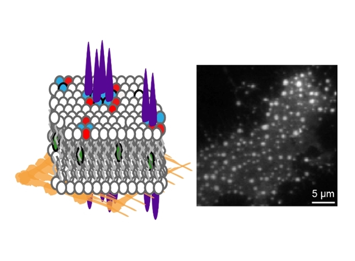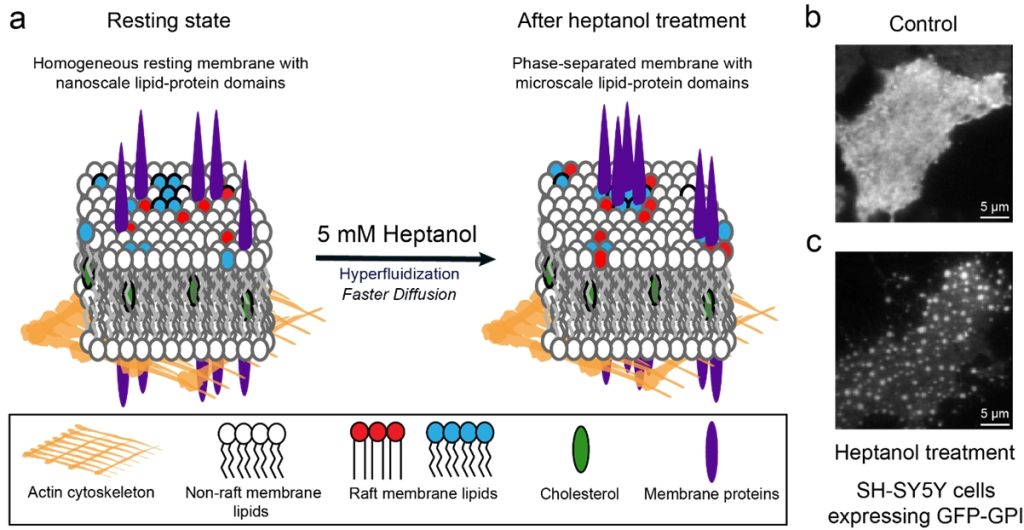
The research team led by Professor Thorsten WOHLAND from the Departments of Biological Sciences and Chemistry, National University of Singapore (NUS) developed a simple fluidizer-based method to determine if a molecule prefers to partition into lipid domains on cell membranes. The team added heptanol to live cells and showed that within 15 minutes it induces clustering of the nanometre-sized lipid domains into larger micrometre-sized domains which are easily detectable by standard fluorescence microscopes. The method works with both molecules that are genetically labelled with fluorescent proteins and those labelled using extrinsic labels, for example, antibodies. Prof Wohland said, “The phase preference of molecules used to be difficult and time-consuming to establish. This new method, detected by chance, provides results in at most 15 minutes on live cells and can essentially be seen by eye in a simple microscope.” The team hopes that this technique will enable a quick and facile identification of domain localisation and will aid the wider research community.

Figure: (a) The schematic shows heptanol-induced membrane fluidization which results in domain clustering. (b) Control: An SH-SY5Y cell in resting state showing homogenous distribution of green fluorescent protein-glycosylphosphatidylinositol (GFP-GPI) on the cell membrane. (c) An SH-SY5Y cell after heptanol treatment showing clustering of GFP-GPI in lipid domains on the cell membrane. [Credit: Journal of Lipid Research]. Read the full story here.
