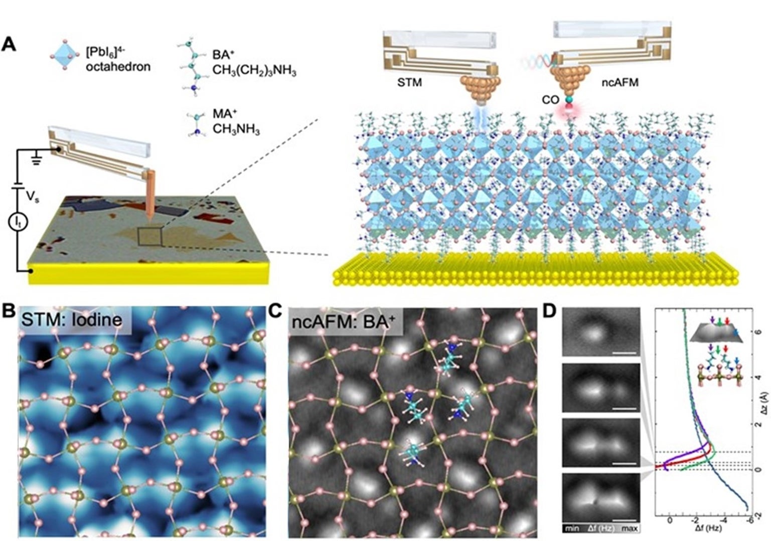
A NUS research team led by Associate Professor Jiong LU, in collaboration with Professor Kian Ping LOH’s research group, both from the Department of Chemistry at the National University of Singapore has developed a method for non-invasive imaging of both the top organic layers and their underlying inorganic lattice in 2D RPP at the sub-angstrom scale. The researchers used a combination of scanning tunnelling microscopy (STM) and imaging techniques (Figure (A)). The STM results provided an atomic reconstruction of the inorganic lead-halide lattice (Figure (B)), while the tip-functionalised ncAFM imaging enabled a visualisation of the top organic layers and its arrangement with respect to the undelaying inorganic lattice at sub-angstrom resolution (Figure (C)). The reconstruction of the on-surface organic layers, presented by a well-ordered array containing pairs of butylammonium cations (Figure (D)), was found to be intimately interlocked with the deformation of the inorganic lattice through hydrogen bonding interactions. This work is jointly undertaken with Prof Pavel JELÍNEK from the Institute of Physics, Czech Academy of Sciences. Prof Lu said, “Our findings not only bring seminal nanoscale insights on the ground state structure of both organic and inorganic motifs in RPPs, but also shed new light on the mechanism of the efficient separation of photoexcited electron–hole pairs and exciton transport in them.”

Figure: (A) Schematics showing the imaging of the RPP surface using a combination of scanning tunnelling microscopy (STM) and tip-functionalised non-contact atomic force microscopy (ncAFM) imaging techniques using a tuning fork–based qPlus sensor. (B) STM image and (C) ncAFM image collected over the same surface area. (D) Δf(Δz) curves acquired over the sites marked by colour-coded arrows in the experimental three-dimensional (3D)-rendered ncAFM image in the inset (top) and side view of the butylammonium pair structure (inset left). [Credit: Science Advances]. Read the full story here.
